This blog is written by our clinicians and aims to keep patients informed with up to date information on medical conditions.
Gynaecological ultrasound: The ins and outs of pelvic ultrasound
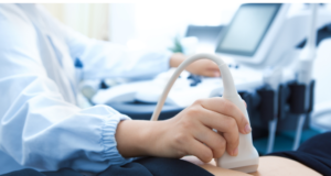
A pelvic ultrasound allows the visualisation of your pelvic organs and structures including the uterus, lining (endometrium), ovaries, cervix and the fallopian tubes. Doppler ultrasound may also show blood flow in certain pelvic organs. Ultrasound uses a special type of camera (transducer) that emits sound waves that reflect off the organs and return back to the transducer, converting the echo into an electronic image of the organs on the ultrasound machine screen.
A pelvic ultrasound can be done either externally (transabdominal) on top of your lower abdomen and as well (with your consent) and internal (transvaginal) examination. The difference between the two is explained below:
For the transabdominal pelvic ultrasound warm gel is placed between the transducer and the skin to allow for smooth movement over the skin and to create a contact medium for the ultrasound soundwaves to travel though. We will ask that you drink adequate water to fill your bladder. Having a full bladder gives clearer pictures for the sonographer performing the examination as it helps to position the uterus and pushes internal bowel gas to the side away from the pelvic organs. The sonographer will move the ultrasound camera around the pelvis to obtain the necessary views of the uterus, ovaries and surrounds. Usually the sonographer can obtain adequate clear images of the uterus, ovaries and other pelvic organs externally however there are a few limiting factors that reduce the image quality and the overall visualisation. The main limiting factor that we face with the transabdominal approach is bowel gas overlying the pelvic organs, this is something that is beyond ultrasound control, sometimes with gentle pressure applied to the lower abdomen the bowel gas will move. If the patient’s bladder is adequality filled it can also reduce the overall visualisation.
Once the transabdominal scan has been completed, the sonographer will offer and inform you about the transvaginal scan (it is optional, but an internal scan gives much clearer pictures).
For the transvaginal pelvic ultrasound (internal scan), you will be asked to empty your bladder and the sonographer uses a special camera/wand that is specially designed for this type of scan. The examination will be explained to the patient and they must provide adequate informed verbal consent for the sonographer to proceed. The look of the camera is very daunting as it is quite long and thin, however ladies do not fear it is mostly handle (to be honest it is much easier and more comfortable than a smear test!). The probe is covered with a special cover and cold gel (sorry girls this gel is cold and cannot be warmed) is applied to the tip. The camera itself is inserted only a short way into the vaginal canal, the camera is slowly moved up and down and from left to right to view the internal pelvic organs and obtain the adequate views and measurements required. The scan itself should not be painful, however you may feel slight pressure and if you are experiencing pain please just inform the sonographer and they will stop or reduce the pressure that may be causing the sudden pain. Everyone’s experience is different and pain levels/tolerances differ, but if you can sit back and relax it will all be over and done within minutes. The scan itself can take up to 5-10 minutes.
In terms of gynaecological ultrasound, the internal (transvaginal) ultrasound is considered to be gold standard; however, as a patient you still have the right to refuse the test for your own personal reasons. The main difference between the external Vs internal ultrasound is the image clarity and the higher resolution images that are obtained when doing the internal pelvic ultrasound, this is simply due to depth and other pitfalls (such as bowel gas). When we scan on top of your abdomen we must account to the subcutaneous adipose tissue and the bladder volume, which can sometimes mean we view the uterus at 5-10cm away, compared to the internal scan where the uterus is often 1-2 cm away. The lining of the uterus (the endometrium) is seen clearly and in detail, the detail which is simply not possible at times externally.
When is it diagnostically important to consider an internal scan:
- An internal ultrasound can provide GP’s and specialist important valuable information when considering IVF treatment, follicular tracking or investigating subfertility.
- If you are being investigated for hormonal imbalances such as polycystic ovaries it is important to have an internal ultrasound (if applicable) to assess the size of the ovaries, follicular count and distribution.
- Internally we are able to obtain images with higher resolution and better visualisation of smaller intrauterine fibroids, small ovarian lesions and intracavity lesions (endometrial lesions such as polyps), especially when determining the fibroids relationship to the endometrial cavity which is important.
- The muscle wall of the uterus is also appreciated in far greater detail internally, we as a sonographer can assess the look of the uterus, the size and also assess the vascular pattern.
- Internally when we apply light pressure to the uterus and ovaries we can assess to see if they move independently with the surrounds, this assessment cannot be done externally
There are times when an internal scan may not necessary or appropriate. For instance if you have never been sexually active it is important to let the sonographer know as in internal scan it not appropriate and they will aim to provide the referring clinician with adequate information with an external scan only. If the patient is seen to have large fibroids and adnexal masses are better seen externally.
What are some reasons/symptoms that we may require or benefit from a gynaecological ultrasound:
- Heavy or painful periods
- Pelvic pain including pain during intercourse
- Assessing the position and location of an IUD
- Bloating
- Infertility
- Irregular or infrequent periods
- Postmenopausal bleeding
- Incidental findings on other imaging modalities such as CT and MRI.
The pelvic ultrasound may not provide your doctor with all the answers to your problems, but it may be very helpful in diagnosis and management.
The benefit of having a gynaecological examination done at ROC health is that we have GP’s on site that can discuss the results with you following the ultrasound appointment and arrange further testing such as blood tests on the day.
Learn more about our Pelvic health physiotherapy service.
Click here to book an appointment